Facet Joint Injections
![]()
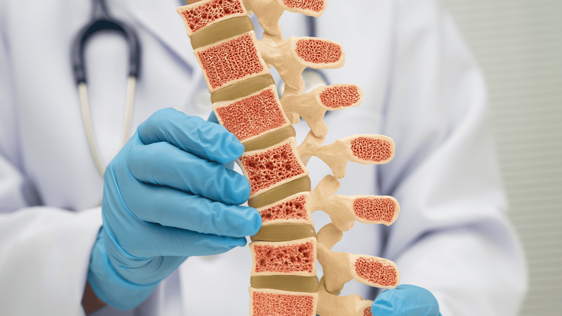
The spine has a repeating sequence of vertebral bones stacked one upon the other connected by discs in the front (anterior) and a pair of facet joints in the back (posterior) situated on the right and left.
Facet joints (also called zygapophysial joints) can become painful due to degenerative changes. Small diameter nerve branches called medial branches transmit the signals from painful facet joints to the central nervous system. Each individual joint surface is serviced by a separate nerve branch, so each facet joint requires two distinct medial branches. There are two injection techniques to determine if a given facet joint(s) is/are contributing to your pain: Medial Branch Block (MBB) Technique or Intraarticular Facet Joint Injection Technique. The MBB numbs the medial branches that service the suspected facet joint(s). After the injection you are given a pain log to take home and compare your pre-injection pain level to what you feel after the medial branch block. The MBB is a “diagnostic” procedure because by itself it is unlikely to deliver lasting facet joint pain relief.
The purpose of the MBB is to determine how much pain can be attributed to the targeted facet joint(s) and to establish how many joints are involved. The intraarticular facet joint injection can provide the same diagnostic information as the MBB, but has the advantage of potentially providing longer term relief of pain symptoms if anti-inflammatory medication is also injected to interrupt the inflammation within the joint(s). During your pre-injection office evaluation Dr. Terebuh will discuss these two facet joint procedure options with you and select the technique that is most appropriate for your particular spine situation and general medical circumstance.
How to Prepare?
![]()
To get ready for your procedure please read the Preparing For Your Spine Injection page of this website.
Before the procedure starts you will speak with Dr. Terebuh in an exam room to answer any final questions you have. He will also verify procedure safety questions. You will then be escorted to the procedure room where all the necessary equipment is located. The procedure table is a rather narrow padded surface and you will be asked to position yourself on your belly. A procedure room assistant will be present to help you if you desire. Positioning pillows can be used to make you as comfortable as possible. The procedure table will elevate several inches and the targeted area of your spine will be sterilized with an alcohol based skin preparation that gives a cool sensation and temporarily tints your skin orange.
A moving x-ray machine called a fluoroscope operated by Dr. Terebuh will be used to guide the entire procedure. The fluoroscope surrounds you like the letter “C” but it will never touch you. Nerves are “invisible” with x-rays so the fluoroscope is used to see the bone landmarks in the spine and increases the comfort, safety, and precision of the injection. The design of the C-arm fluoroscope allows Dr. Terebuh to get the ideal view angle to perform your procedure. The fluoroscope does deliver a dose of radiation similar to an x-ray and the machine settings are adjusted to administer the absolute minimum radiation dose possible for your procedure.
The skin over the targeted landmark will be injected with numbing medicine, which causes a very temporary sensation like a “bee sting”. Most patients agree that the numbing of the skin is the most unpleasant part of the entire procedure (fortunately it is very brief) and the rest of the process feels like movement and pressure rather than pain.
Each needle movement is verified by the fluoroscope to ensure your safety and your comfort. Once the needle tip is in the ideal position relative to the bone landmarks, a small amount of x-ray dye (contrast) is injected. This dye is visible on the fluoroscope and Dr. Terebuh uses it to verify that the flow of the dye goes into the facet joint and to make sure blood vessels are not carrying it away from the target. (If you have an X-ray dye allergy, this verification step with the x-ray dye cannot take place.) The final step is slowly injecting the treatment medication into the facet joint precisely to the desired location. In the case of a MBB, only numbing medicine is injected into specific locations to block the appropriate medial branches (shown in videos below). A bandage will be applied to the injection site and you be escorted back to the original exam room for the after injection observation, monitoring, and discharge process.

Medial Branch Brock
FIND OUT

Medial Branch Block Cervical
FIND OUT

Anatomy of the Spine
FIND OUT